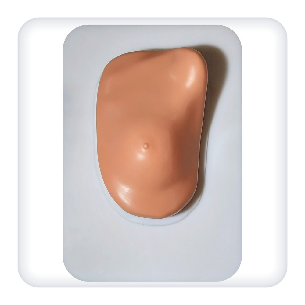
The simulator is a model of a woman’s torso fragment, including the mammary gland, based on a stand. The material of the model is close to its natural values in terms of its physical and ultrasonic properties.
Precise ultrasound anatomy, including pathologies and neoplasms:
- imitation of malignant and benign tumors;
- a cyst;
- Cooper’s ligaments;
- premammary and retromammary fiber;
- glandular tissue.
- mammary gland duct, muscle tissue;
- axillary lymph nodes.
Skills developed on the simulator:
- full ultrasound scan of the mammary gland with real equipment;
- ultrasound visualization of key anatomical landmarks;
- identification of mammary gland ducts;
- visualization of pathological formations;
- detection and measurement of cysts and tumors.
The simulator is intended for use in medical educational institutions.
Equipment:
Simulator for practicing mammary gland ultrasound skills
Documentation:
- Product data sheet
- User Manual
Material:
Polyurethane, Silicone, plastic


